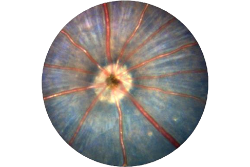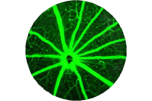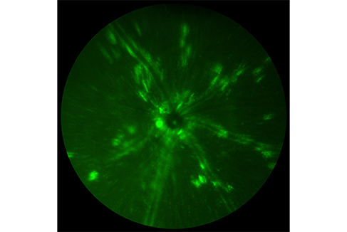



High-resolution Fundus Imaging System
Accurately records fundus structures to empower ophthalmic disease research
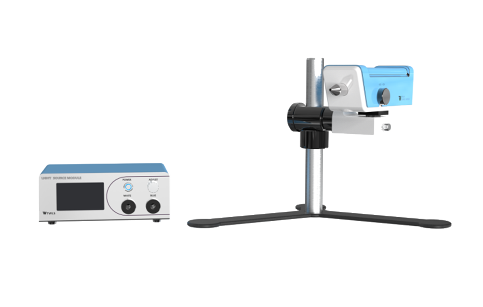
Accurately records fundus structures to empower ophthalmic disease research
1. High-definition Fundus Imaging
Captures clear images of the retina, blood vessels and optic disc to enable intuitive observation and recording of structural changes in the fundus.
2. Non-contact Detection
Adopts safe optical imaging method. No tissue processing required for fast and non-invasive fundus image acquisition.
3. Disease Model Evaluation
Supports animal model studies of diabetic retinopathy, optic nerve injury, retinal degenerative diseases and more.
4. Intelligent Image Analysis
Equipped with image processing software to realize quantitative analysis of vascular parameters, lesion areas and image data.
We will customize professional medical research solutions for you
Contact Us